Cells that make up human tissues. Nervous tissue. Nerve tissue consists of nerve cells with their processes and the endings of these processes. Tests and assignments
Extract from the work program on the topic “Cell. Fabrics"
|
Theory |
Practice | |
|
2 hours |
2 hours 2 hours |
Cell. Fabrics. Cell structure and functions. Concept of fabric. Types of fabrics. Representation cell as a structural unit with the properties of a living histological features of various types of tissues Knowledge structure of the cell, its structures, functions of the nucleus, cell membrane, cytoplasm, organelles cell life cycle, types of cell division properties of a cell as an elementary unit of living things fabric - definition, classification features of the structure and topography of epithelial, connective, muscle and nervous tissues, their types functional significance of different types of tissues Skills be able to distinguish cells and intercellular substance under a microscope be able to distinguish between different types of epithelial, connective, muscle tissue be able to distinguish its structures in a cell, indicating the features of their structure and function be able to give a brief morphological and functional characteristics of tissues |
Lecture topic: "Cell.Tissue"
Cell is the smallest structural one that has all the characteristics of a living thing.
Living things are characterized by a number of properties:
The ability to self-reproduce;
Variability;
Metabolism;
Irritability;
Adaptation.
The combination of these properties is first discovered at the cellular level.
Cell is an ordered structural system of biopolymers bounded by an active membrane. It is a microscopic formation, varying in size and shape.
Cells were discovered and described more than 300 years ago. Robert Hooke observed plant cells using magnifying lenses. Cytology (the science of cells) received its greatest development after T. Schwann (1838) formulated the cell theory, combining all existing research results. Currently, cell theory is based on the following basic principles:
cell is the smallest unit of living things;
cells of different organisms are similar in structure and function (homologous);
Cell reproduction occurs by dividing the original cell.
cells are part of a multicellular organism, where they are united into tissues and organs and are connected by intercellular, humoral and neural forms of regulation.
According to the second principle of the theory, cells of various organisms, despite their diversity, have common structural principles. Each cell consists of a plasmalemma (membrane), cytoplasm and most cells - a nucleus.
Let's look at the characteristics of the cell components.
Plasmolemma is a membrane structure (a thin layer consisting of a double layer of lipids connected to proteins) and performs barrier transport and receptor functions. It separates the cytoplasm of the cell from the external environment. The transport function of the plasmalemma is carried out by various mechanisms. Exists passive transfer molecules by diffusion (ions), osmosis (water molecules), active transfer – with the expenditure of energy ATP and with the help of enzymes – permeases (transfer of AA, sodium, sugars). The transfer of larger molecules is called endocytosis. Its main varieties are phagocytosis – transfer of solid particles and pinocytosis – transfer in liquid media. Particles captured by the cell are immersed, surrounded by a portion of the cytoplasm (phagosomes and pinosomes) and merge with lysosomes, which subject them to cleavage. The receptor function of the plasmalemma is the “recognition” by the cell of various chemical (hormones, proteins) and physical (light, sound) factors with the help of receptors located in the plasmalemma (polysaccharides, glycoproteins).
The plasmalemma can form poisonous special formations - microvilli, brush border, cilia and flagella, as well as various intercellular contacts.
Microvilli – outgrowths of the cytoplasm limited by the plasmalemma (many in the epithelial cells of the intestine and kidneys); increase cell surface area.
Cilia and flagella - outgrowths of the cytoplasm, the origin of which is associated with centrioles, serve as an apparatus for cell movement.
Intercellular contacts – plasmalemma structures that ensure the connection and interaction of cells (transfer of ions, molecules).
Cytoplasm consists of hyaloplasm and organelles and inclusions located in it.
Hyaloplasma - the internal environment of the cell, a structureless, translucent, semi-liquid formation, capable of changing its function. state. It contains proteins and enzymes, transp. RNA, amino acids, polysaccharides, ATP, various ions. The main function is to ensure the chemical interaction of the structures located in it.
Organelles divided into membrane and non-membrane.
Membrane ones include: endoplasmic reticulum
mitochondria
App. Golgi
lysosomes
Non-membrane ones include: ribosomes
polysomes
microtubules
centrioles
EPS – a system of tubes, cisterns, vacuoles bounded by one membrane. There are granular and agranular EPS. Granular is characterized by the presence of granules - ribosomes.
The main function of the EPS is the synthesis of substances and their transport to various parts of the cell and to the external environment. In the agranular EPS, lipids and carbohydrates are synthesized, and in the granular EPS, proteins are synthesized.
Mitochondria – round or rod-shaped structures formed by two membranes (outer and inner, which forms outgrowths inward - cristae, immersed in a matrix in which ribosomes and granules are located). ATP is formed at the cristae. The main function of mitochondria is to ensure cellular respiration and process ATP, the energy of which is used for cell movement, muscle contraction, processes of synthesis and secretion of substances, and the passage of substances through membranes.
Golgi complex – multiple and single dictyosomes (membrane structures consisting of cisterns with expansions, small transport vesicles, large secretory vesicles and granules). The Golgi complex is involved in the secretion process (proteins synthesized in the ER ribosomes enter the Golgi complex), synthesizes polysaccharides, and forms lysosomes.
Lysosomes – these are small bubbles 0.2 - 0.4 microns in size, bounded by a single membrane and containing more than 40 different enzymes that break down proteins, nucleic acids, lipids, and carbohydrates. The function of lysosomes is to digest various substances coming from outside and destroy aging or defective structures in the cell itself.
Non-membrane organelles:
Ribosomes – Protein synthesis organelles are formed in the nucleolus. They consist of two subunits - small and large, each of which is built from a twisted strand of ribonucleoprotein, where proteins and ribosomal RNA are equally represented. Young cells are characterized by the presence of free ribosomes, which provide protein synthesis for the cell itself (growth). In differentiated cells, the number of ribosomes and polysomes associated with the EPS and ensuring the synthesis of proteins “for export” (cell secretion) increases.
Microtubules – hollow cylinders with a diameter of 24 nm, consisting of the protein tubulin. They can constantly form in the hyaloplasm, participating in the formation of the cell cytoskeleton. They are part of centrioles, cilia, flagella, and spindles.
Centrioles – lie in pairs, each consisting of microtubules. They are located perpendicular to each other and are surrounded by radially extending microtubules (centrosphere)
Microfilaments and microfibrils – performs supporting-framework and contractile functions in the cell, which ensures cell movement and movement of organelles and inclusions in the hyaloplasm.
Core performs the most important functions in the cell - storage and transmission of genetic information and ensuring protein synthesis (formation of all types of RNA - inf., transsp., ribosomal., synthesis of ribosomal proteins). Protein structure and function change during the cell cycle—the time from division to division or from division to death.
The nucleus of an interphase cell (non-dividing) consists of a nuclear membrane, chromatin, nucleolus and karyoplasm (nucleoplasm)
Nuclear envelope consists of two membranes - outer and inner. The shell contains pores (complexes) that ensure the passage of macromolecules from the nucleus into the cytoplasm. One of the functions of the nuclear membrane is to fix chromosomes and ensure their spatial position.
Chromosomes are constantly present in the nucleus and are clearly visible only during mitosis. In the interphase nucleus, chromosomes are dispersed and not visible. Consists of DNA, protein, RNA.
nucleolus - a round body in which ribosomes are formed. The number of nucleoli varies in different cells. An increase in the number and size of nucleoli indicates a high intensity of RNA and protein synthesis.
Cell life cycle
The cell, being part of an integral multicellular organism, performs functions characteristic of living things. These include reproduction.
The main form of cell reproduction is mitosis (indirect division). Mitosis consists of 4 main phases: prophase, metaphase, anaphase, telophase.
- prophase Chromosomes condense, they become visible, each chromosome consists of two sister chromosomes - chromatids, nucleoli become smaller and disappear, the nuclear shell is destroyed, the number of ribosomes and grains decreases. The EPS breaks up into small vacuoles, the centrioles diverge, and the spindle begins to form (microtubules extending from the centrioles);
- metaphase the spindle is completed to form and the chromosomes are located on the equatorial plane of the cell;
- anaphase halves of chromosomes lose connection in the region. centromeres and diverge to the poles of the cell, a diploid set of chromosomes (46 in humans) goes to the pole;
- telophase the structures of the interphase nucleus are restored - despiralization of chromosomes, reconstruction of the nuclear membrane, appearance of nucleoli, division of the cell body into two parts.
The duration of mitosis and its individual phases varies in different cells from 30 minutes. Up to 3 hours or more (interphase 10-30 hours, prophase 30-60 hours, metaphase 2-10 minutes, anaphase 2-3 minutes, telophase 20-30 minutes). The number of mitoses in tissues and organs is an indicator of the intensity of their growth and regeneration (physiological and regenerative) in normal and pathological conditions.
A type of mitosis is meiosis - the division of maturing germ cells, which leads to a halving of the number of chromosomes, i.e. formation of the haploid number of chromosomes (23 in humans). Meiosis consists of two successive divisions with a short interphase - reduction (the number of chromosomes is reduced) and evolution (mitosis).
In addition to the ability to reproduce, a cell has a number of properties that characterize living things:
Metabolism From the external environment (blood, lymph, tissue fluid), substances that go towards building the cell and oxidative processes enter through the semi-permeable membrane; waste products of the cell are removed through the membrane.
Permeability cells depends on various factors, including from
salt concentrations Substances can enter through phagocytosis
and pinocytosis.
Secretion- substances secreted by cells (hormones,
enzymes, biologically active substances).
Irritability ability to respond with specific reactions to
influence of an external stimulus. Muscle, nerve, and glandular cells have the highest degree of irritability -
excitability. As a particular type of irritability is the ability of cells to move - leukocytes, macrophages, fibroblasts, sperm.
Fabrics. Species, their morphological and functional characteristics.
There are 4 types of tissues in the human body:
epithelial;
connecting;
muscular;
Epithelium covers the surfaces of the body, mucous and serous membranes of internal organs and forms most of the glands.
Covering epithelium does:
barrier function
exchange function
protective function
Glandular epithelium performs a secretory function.
General characteristics of the integumentary epithelium.
Variety of morphological forms;
There is no intercellular substance;
The cells are arranged in a layer;
Located on the basement membrane;
There are no blood vessels;
High regeneration.
Structure and functions of the integumentary epithelium.
Morphological classification of epithelium:
Single layer epithelium-
Cubic
Prismatic
Multi-row
Stratified epithelium
Non-keratinizing
keratinizing
Transition
Glandular epithelium.
Glands (gianduiae) perform a secretory function and are derivatives of the glandular epithelium.
Many glands are independent organs (pancreas, thyroid), other glands are part of an organ (stomach glands).
All glands are divided into:
Endocrine, producing their secretions (hormones) into the blood.
Exocrine cells produce secretions into the external environment (on the skin and in organ cavities).
According to their structure, exocrine glands are divided into simple and complex with branching excretory ducts. According to the chemical composition of the secretion, they are divided into protein (serous), mucous, and protein-mucous.
Support-trophic tissues.
This group includes blood and lymph, as well as connective tissue. They all have a similar structure: they contain a well-developed intercellular substance. All tissues of this group perform a trophic function (blood, lymph) and a supporting function (cartilage, bone).
Blood, lymph, loose connective tissue make up internal environment of the body.
Connective tissue.
This group includes:
connective tissue itself(loose and dense)
with special properties(reticular, fatty, mucous, pigmented)
skeletal connective tissue(cartilage, bone tissue)
Connective tissue is characterized by a variety of cells and a well-developed intercellular substance, consisting of fibers and a basic amorphous substance. The classification is based on the ratio of cells and intercellular substance, as well as the degree of orderliness in the arrangement of fibers.
Tissue cells : fibroblasts, macrophages, plasmacytes, mast cells, adipocytes, pigmentocytes, adventitial cells, blood leukocytes.
Intercellular substance : consists of collagen, reticular, elastic fibers and ground substance.
Loose fibrous connective tissue - accompanies blood and lymphatic vessels, forms the stroma of many organs.
Dense fibrous connective tissue contains a large number of densely located fibers and a small number of cellular elements. This tissue underlies tendons, ligaments, and fibrous membranes.
Cartilage tissue consists of cells (chondrocytes) and a large amount of intercellular substance.
There are three types of cartilage tissue:
hyaline (fetal skeleton, costosternal junction, laryngeal cartilages, articular surfaces)
elastic (at the base of the auricle)
fibrous (intervertebral discs, semi-movable joints)
Bone a specialized type of connective tissue with highly mineralized intercellular substance containing about 70% inorganic substances (calcium phosphates).
There are two types of bone tissue - reticulofibrous and lamellar.
Bone cells include: osteocytes, osteoblasts, osteoclasts.
Lamellar bone tissue the most common bone tissue in the adult body. It consists of bone plates formed by bone cells and a mineralized ground substance with collagen fibers. In adjacent plates, the fibers have different directions, which achieves greater strength of the bone tissue. The compact and spongy bones of the skeleton are built from this tissue.
Muscle.
Provides movement in space of the body as a whole and its parts. Muscle tissue has the ability to contract under the influence of nerve impulses, which is accompanied by a change in membrane potentials. Contraction occurs due to the content of myofibrils in muscle cells, due to the interaction of the actin and myosin proteins with the participation of Ca ions.
All muscle tissues are divided into two subgroups:
smooth muscle tissue (threads of actin and myosin myofibrils do not have transverse striations) are present on the walls of internal organs and have greater extensibility and less excitability than skeletal tissue;
striated tissues (actin and myosin myofibrils create cross-striations) form cardiac muscle tissue and skeletal muscle tissue.
Nervous tissue.
Nervous tissue regulates the activity of tissues and organs, their relationship and connection with the environment. Nervous tissue consists of neurons (nerve cells) and neuroglia, which perform supporting, trophic, delimiting and protective functions.
Neurons conduct nerve impulses from the point of origin to the working organ. Each cell has processes - axon(conducts an impulse from the cell body and ends on a neighboring neuron, muscle, gland) and dendrite(carries impulse to the body, there can be several of them and they branch). Neurons are divided according to the number of processes:
Unipolar (1 branch)
Bipolar (2 branches)
Multipolar (3 or more processes)
Bipolar cells also include pseudounipolar cells (the axon and dendrite of these cells begin with a common outgrowth). The processes of nerve cells, usually covered with membranes, are called nerve fibers. All nerve fibers end in terminal apparatuses, which are called nerve endings, they are divided into three groups
Effector (motor and secretory)
Receptor (sensitive)
Terminal (interneuron synapses).
Epithelial tissue
Epithelial (integumentary) tissue, or epithelium, is a boundary layer of cells that lines the integument of the body, the mucous membranes of all internal organs and cavities, and also forms the basis of many glands.

The epithelium separates the organism (internal environment) from the external environment, but at the same time serves as an intermediary in the interaction of the organism with the environment.
Epithelial cells are tightly connected to each other and form a mechanical barrier that prevents the penetration of microorganisms and foreign substances into the body.
Epithelial tissue cells live for a short time and are quickly replaced by new ones (this process is called regeneration).

Epithelial tissue is also involved in many other functions: secretion (exocrine and endocrine glands), absorption (intestinal epithelium), gas exchange (lung epithelium).
The main feature of the epithelium is that it consists of a continuous layer of tightly adjacent cells. The epithelium can be in the form of a layer of cells lining all surfaces of the body, and in the form of large accumulations of cells - glands: liver, pancreas, thyroid, salivary glands, etc. In the first case, it lies on the basement membrane, which separates the epithelium from the underlying connective tissue . However, there are exceptions: epithelial cells in the lymphatic tissue alternate with connective tissue elements; such epithelium is called atypical.
Epithelial cells, arranged in a layer, can lie in many layers (stratified epithelium) or in one layer (single-layer epithelium). Based on the height of the cells, epithelia are divided into flat, cubic, prismatic, and cylindrical.
Connective tissue

Consists of cells, intercellular substance and connective tissue fibers. It consists of bones, cartilage, tendons, ligaments, blood, fat, it is present in all organs (loose connective tissue) in the form of the so-called stroma (framework) of organs.
In contrast to epithelial tissue, in all types of connective tissue (except adipose tissue), the intercellular substance predominates over the cells in volume, i.e. the intercellular substance is very well expressed. The chemical composition and physical properties of the intercellular substance are very diverse in different types of connective tissue. For example, blood - the cells in it “float” and move freely, since the intercellular substance is well developed.
In general, connective tissue makes up what is called the internal environment of the body. It is very diverse and is represented by various types - from dense and loose forms to blood and lymph, the cells of which are in the liquid. The fundamental differences in the types of connective tissue are determined by the ratios of cellular components and the nature of the intercellular substance.
IN dense Fibrous connective tissue (muscle tendons, joint ligaments) is dominated by fibrous structures and experiences significant mechanical loads.
Loose fibrous connective tissue is extremely common in the body. It is very rich, on the contrary, in cellular forms of different types. Some of them are involved in the formation of tissue fibers (fibroblasts), others, which is especially important, provide primarily protective and regulatory processes, including through immune mechanisms (macrophages, lymphocytes, tissue basophils, plasma cells).
Bone

The bone tissue that forms the bones of the skeleton is very strong. It maintains body shape (constitution) and protects organs located in the skull, chest and pelvic cavities, and participates in mineral metabolism. The tissue consists of cells (osteocytes) and intercellular substance in which nutrient channels with blood vessels are located. The intercellular substance contains up to 70% mineral salts (calcium, phosphorus and magnesium).
In its development, bone tissue passes through fibrous and lamellar stages. In various parts of the bone it is organized in the form of compact or spongy bone substance.
Cartilage tissue

Cartilage tissue consists of cells (chondrocytes) and intercellular substance (cartilage matrix), characterized by increased elasticity. It performs a supporting function, as it forms the bulk of cartilage.
There are three types of cartilage tissue: hyaline, which is part of the cartilage of the trachea, bronchi, ends of the ribs, and articular surfaces of bones; elastic, forming the auricle and epiglottis; fibrous, located in the intervertebral discs and joints of the pubic bones.
Adipose tissue

Adipose tissue is similar to loose connective tissue. The cells are large and filled with fat. Adipose tissue performs nutritional, shape-forming and thermoregulatory functions. Adipose tissue is divided into two types: white and brown. In humans, white adipose tissue predominates, part of it surrounds the organs, maintaining their position in the human body and other functions. The amount of brown adipose tissue in humans is small (it is found mainly in newborns). The main function of brown adipose tissue is heat production. Brown adipose tissue maintains the body temperature of animals during hibernation and the temperature of newborns.
Muscle
Muscle cells are called muscle fibers because they are constantly stretched in one direction.

Classification of muscle tissue is carried out on the basis of the structure of the tissue (histologically): by the presence or absence of transverse striations, and on the basis of the mechanism of contraction - voluntary (as in skeletal muscle) or involuntary (smooth or cardiac muscle).
Muscle tissue has excitability and the ability to actively contract under the influence of the nervous system and certain substances. Microscopic differences allow us to distinguish two types of this tissue - smooth (unstriated) and striated (striated).

Smooth muscle tissue has a cellular structure. It forms the muscular membranes of the walls of internal organs (intestines, uterus, bladder, etc.), blood and lymphatic vessels; its contraction occurs involuntarily.

Striated muscle tissue consists of muscle fibers, each of which is represented by many thousands of cells, fused, in addition to their nuclei, into one structure. It forms skeletal muscles. We can shorten them at will.
A type of striated muscle tissue is cardiac muscle, which has unique abilities. During life (about 70 years), the heart muscle contracts more than 2.5 million times. No other fabric has such strength potential. Cardiac muscle tissue has transverse striations. However, unlike skeletal muscle, there are special areas where the muscle fibers meet. Thanks to this structure, the contraction of one fiber is quickly transmitted to neighboring ones.
This ensures simultaneous contraction of large areas of the heart muscle.
Nervous tissue
Nervous tissue consists of two types of cells: nerve (neurons) and glial. Glial cells are closely adjacent to the neuron, performing supporting, nutritional, secretory and protective functions.

Neuron is the basic structural and functional unit of nervous tissue. Its main feature is the ability to generate nerve impulses and transmit excitation to other neurons or muscle and glandular cells of working organs. Neurons can consist of a body and processes. Nerve cells are designed to conduct nerve impulses. Having received information on one part of the surface, the neuron very quickly transmits it to another part of its surface. Since the processes of a neuron are very long, information is transmitted over long distances. Most neurons have processes of two types: short, thick, branching near the body - dendrites and long (up to 1.5 m), thin and branching only at the very end - axons. Axons form nerve fibers.
A nerve impulse is an electrical wave traveling at high speed along a nerve fiber.
Depending on the functions performed and structural features, all nerve cells are divided into three types: sensory, motor (executive) and intercalary. Motor fibers running as part of nerves transmit signals to muscles and glands, sensory fibers transmit information about the state of organs to the central nervous system.
Human tissues
| Fabric group | Types of fabrics | Tissue structure | Location | Functions |
| Epithelium | Flat | The surface of the cells is smooth. Cells are tightly adjacent to each other | Skin surface, oral cavity, esophagus, alveoli, nephron capsules | Integumentary, protective, excretory (gas exchange, urine excretion) |
| Glandular | Glandular cells produce secretions | Skin glands, stomach, intestines, endocrine glands, salivary glands | Excretory (secretion of sweat, tears), secretory (formation of saliva, gastric and intestinal juice, hormones) | |
| Ciliated (ciliated) | Consists of cells with numerous hairs (cilia) | Airways | Protective (cilia trap and remove dust particles) | |
| Connective | Dense fibrous | Groups of fibrous, tightly packed cells without intercellular substance | The skin itself, tendons, ligaments, membranes of blood vessels, cornea of the eye | Integumentary, protective, motor |
| Loose fibrous | Loosely arranged fibrous cells intertwined with each other. The intercellular substance is structureless | Subcutaneous fatty tissue, pericardial sac, nervous system pathways | Connects skin to muscles, supports organs in the body, fills gaps between organs. Provides thermoregulation of the body | |
| Cartilaginous | Living round or oval cells lying in capsules, the intercellular substance is dense, elastic, transparent | Intervertebral discs, laryngeal cartilage, trachea, auricle, joint surface | Smoothing the rubbing surfaces of bones. Protection against deformation of the respiratory tract and ears | |
| Bone | Living cells with long processes, interconnected, intercellular substance - inorganic salts and ossein protein | Skeleton bones | Supportive, motor, protective | |
| Blood and lymph | Liquid connective tissue consists of formed elements (cells) and plasma (liquid with organic and mineral substances dissolved in it - serum and fibrinogen protein) | Circulatory system of the whole body | Carries O2 and nutrients throughout the body. Collects CO2 and dissimilation products. Ensures the constancy of the internal environment, chemical and gas composition of the body. Protective (immunity). Regulatory (humoral) | |
| Muscular | Cross-striped | Multinucleate cylindrical cells up to 10 cm in length, striated with transverse stripes | Skeletal muscles, cardiac muscle | Voluntary movements of the body and its parts, facial expressions, speech. Involuntary contractions (automatic) of the heart muscle to push blood through the chambers of the heart. Has excitability and contractility properties |
| Smooth | Mononuclear cells up to 0.5 mm long with pointed ends | Walls of the digestive tract, blood and lymph vessels, skin muscles | Involuntary contractions of the walls of internal hollow organs. Raising hair on the skin | |
| Nervous | Nerve cells (neurons) | Nerve cell bodies, varied in shape and size, up to 0.1 mm in diameter | Forms the gray matter of the brain and spinal cord | Higher nervous activity. Communication of the organism with the external environment. Centers of conditioned and unconditioned reflexes. Nervous tissue has the properties of excitability and conductivity |
| Short processes of neurons - tree-branching dendrites | Connect with processes of neighboring cells | They transmit the excitation of one neuron to another, establishing a connection between all organs of the body | ||
| Nerve fibers - axons (neurites) - long processes of neurons up to 1 m in length. Organs end with branched nerve endings | Nerves of the peripheral nervous system that innervate all organs of the body | Pathways of the nervous system. They transmit excitation from the nerve cell to the periphery via centrifugal neurons; from receptors (innervated organs) - to the nerve cell along centripetal neurons. Interneurons transmit excitation from centripetal (sensitive) neurons to centrifugal (motor) neurons |
Nerve tissue consists of nerve cells with their processes and the endings of these processes. It also includes formations that have a supporting and trophic significance for the nervous tissue itself and are called neuroglia /macro- And microglia/.
Each nerve cell contains cytoplasm and a round nucleus
cell processes, since the neuron body forms a protrusion corresponding to each process.
The nature of the origin of the processes makes it possible to differentiate neurons to unipolar(with one shoot, T-shaped, divided into two branches), bipolar(with two shoots
Rice. 9. Structure of the motor
neuron:
Perikaryon; 2 - axon and nerve fiber; 3 - nerve endings in the muscle; 4 - dendrites; 5 - pulpy shell; 6 - Ranvier interceptions. The diagram compares light and electron microscopy (according to G.F. Ivanov and Kovalsky, modified)
or slightly oval in shape. Mitochondria and the Golgi complex are clearly visible in the cytoplasm. There are tigroid deposits that arise from modifications of the endoplasmic reticulum. Under an electron microscope, neurofilaments are determined - threads about 10 nm thick.
A nerve cell with its processes is called neuron(Fig. 9). It is a structural unit of nervous tissue. Nerve cells whose processes go to organs (for example, muscles) and non-
kami) and multipolar(multi-processed).
Some processes of nerve cells - short, protoplasmic, tree-like branching - dendrites; others are long neurites, or axons. The length of the processes of nerve cells can be very long (in some places more than 1 m). Along neurites, stimuli travel from the cell body, while through dendrites - to the cell body.
The processes continue as part of the nerve fibers in the form cylinder axle cylinders, usually covered with glial membranes of simpler or
Main content.
- Classification of mechanical fabrics.
- Characteristics of collenchyma.
- Characteristics of sclerenchyma.
You probably know that plants do not have a skeleton that would help it withstand the weight of its own organs (branches, leaves, flowers, fruits, etc.), as well as the action of wind, rain, snow, etc. In addition, due to the lack of a nervous system, it cannot quickly mobilize (as animals do) muscular resistance to storm gusts and various other pressures and loads. However, the plant successfully withstands these loads. The strength it needs is provided by the complex of all tissues. A plant as a whole can be considered as a natural structure that exists thanks to the expedient principles of its structure.
The significant strength of the plant is achieved, first of all, by the turgor (fluid pressure inside the cell equal to the pressure on the cell outside) of living cells and tissues.
Play a major role in the strength of plants mechanical fabrics. 
Higher plants spend their entire lives in one place. The body shape of plants, the characteristics of trees, represents a significant surface area. The equipment of this surface in the form of a powerful crown, as well as a deep root system, must resist storms, downpours, hail, etc. over tens and hundreds of years of life. The pressure of the crown itself on the tree trunk is a constant force, the tension of which varies depending on the number of fruits, branches, leaves, snow, etc.
It is usually believed that only special mechanical tissues perform the function of resisting all types of pressure and load. Meanwhile, the strength of plant organs depends not only on mechanical tissues, but also on the entire mass of other tissues.
The plant can be compared in many cases to a reinforced concrete structure. The latter consists of an iron frame (reinforcement), around and in the spaces of which the main mass (concrete) is placed. The frame, immersed in a mass of concrete, plays the role of a skeleton, a binder of concrete materials. The complex of mechanical tissues (plant reinforcement) is similar to a frame located in the mass of the main, conductive and other tissues.
There are mechanical tissues with uniformly and unevenly thickened cell walls.
Classification of mechanical fabrics

 Collenchyma.
Tissue whose cells have unevenly thickened cells. There are angular and lamellar collenchyma. Collenchyma cell walls consist of cellulose, hemicellulose and pectin substances. The cells are chlorophyll-bearing, so collenchyma is not found in underground organs.
Collenchyma.
Tissue whose cells have unevenly thickened cells. There are angular and lamellar collenchyma. Collenchyma cell walls consist of cellulose, hemicellulose and pectin substances. The cells are chlorophyll-bearing, so collenchyma is not found in underground organs.

Collenchyma in plantain leaves
In many ways, collenchyma resembles parenchyma, but it is characterized by additional deposition of cellulose in the corners of cells. This deposition occurs after the formation of the primary cell wall. In addition, collenchyma cells extend parallel to the long axis of the organ in which this tissue is embedded. In stems and leaf petioles, the supporting function of collenchyma is also enhanced due to the fact that this tissue is located near the surface of the organ. Often it lies directly under the epidermis, in the outer zone of the cortex, gradually passing into the parenchyma to the central part of the organ, i.e. forms in three dimensions as if into a hollow cylinder. In other cases, it can form ribs that increase the strength of the organ, as in the fleshy petioles of celery leaves or the ribbed stems of plants such as damselfish. In dicotyledonous leaves, collenchyma surrounds the midrib and serves as a support for vascular bundles.
 Cell collenchyma angularis has the shape of a hexagonal polyhedron, in which the thickening of the cellulose membrane runs along the ribs, and in the cross section, the thickening of the cell wall is noticeable at the corners of this polyhedron. Angular collenchyma is found in the stems of dicotyledonous plants (mostly herbaceous), in leaf petioles on both sides of large leaf veins. Collenchyma does not interfere with the lengthwise growth of the organ in which it is located.
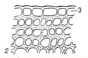 Cell lamellar collenchyma has the shape of a parallelepiped, in which only a pair of faces (walls) are thickened, visible in the cross section from the tangential sides, i.e. parallel to the surface of the stem. Lamellar collenchyma is found, as a rule, in the stems of woody plants.
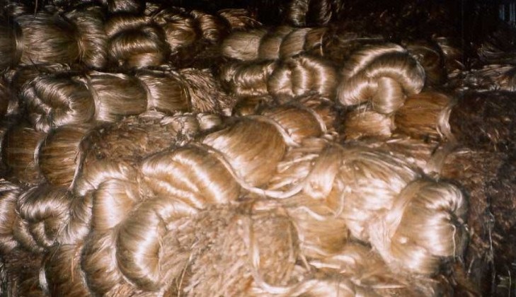
Sclerenchyma. Tissue whose cells are lignified (impregnated with lignin, a special substance that causes lignification)

1 – 6 – sclerenchyma fibers 7-8 – sclereids
uniformly thickened cell walls. The nucleus and cytoplasm are destroyed. There are two types of sclerenchyma - sclerenchyma fibers and sclereids. The fibers are collected in bundles or strands.

Coconut fiber
Sclerenchyma fibers form tissue consisting of elongated cells with pointed ends and pore channels in the cell walls. These cells are tightly adjacent to each other and their membranes are highly durable. In a cross section, the cells are multifaceted.
When sclerenchyma fibers occur in wood (xylem), they are called wood fibers (libriform). They protect blood vessels from pressure from other tissues, being

Hemp - hemp fiber
mechanical part of xylem (wood).
If sclerenchyma fibers are found in the phloem, then they are called bast fibers(cambiform). Bast fibers can also be non-lignified, while possessing great strength and elasticity, which is of great practical use in the textile and other industries (for example, flax, jute, hemp fibers).
 |
 |
 |
|
Hemp (hemp fiber) |
Lapti (linden bast) |
Bask tues |
 |
 |
 |
|
Flax fiber |
Jute fiber |

Sclereids (stony cells)
Sclereids usually arise from the cells of the main parenchyma as a result of thickening and lignification (lignification) of their cell walls. They have different shapes and are found in many plant organs. Sclereids of more or less isodiametric shape (with the same cell diameter) are called brachysclereids, or stony cells (in pear fruits, cherry pits)
Sclereids that have expansion at both ends of the cell - osteosclereids - are found in tea leaves. Sclereids whose shape resembles a star are called astrosclereids (in camellia leaves). Elongated rod-shaped sclereid cells are found in legume seeds.
The shell of nuts is also formed by sclereids.

 |
 |
 |
There are four main types of tissue: epithelial, connective, muscle and nervous.
Epithelial tissue consists of cells that fit very tightly together. The intercellular substance is poorly developed. Epithelial tissue covers the surface of the body from the outside (skin), and also lines the inside of hollow organs (stomach, intestines, renal tubules, pulmonary vesicles). The epithelium can be single-layered or multilayered. Epithelial tissues perform protective, excretory and metabolic functions.
The protective function of the epithelium is to protect the body from damage and penetration of pathogens. Epithelial tissues include ciliated epithelium, the cells of which on the outer surface have cilia that can move. Through the movement of cilia, the epithelium directs foreign particles outside the body. The ciliated epithelium lines the inner surface of the respiratory tract and removes dust particles that enter the lungs with air.
The excretory function is carried out by the glandular epithelium, the cells of which are capable of forming fluids - secretions: saliva, gastric and intestinal juices, sweat, tears, etc.
The metabolic function of epithelial tissues is to carry out the exchange of substances between the external and internal environment:
release of carbon dioxide and absorption of oxygen in the lungs, absorption of nutrients from the intestines into the blood.
Most epithelial cells die and desquamate during their life (in the skin, digestive tract), so their number must be constantly restored through division.
Connective tissue. This name unites a group of tissues with a common origin and function, but with different structures. The functions of connective tissue are to give strength to the body and organs, maintain and connect all cells, tissues and organs of the body. Connective tissue consists of cells and the main, or intercellular, substance, which can be in the form of fibers or be continuous, homogeneous. Connective tissue fibers are built from the proteins collagen, elastin, etc. The following types of connective tissue are distinguished: dense, cartilaginous, bone, loose and blood. Dense connective tissue is found in the skin, tendons, and ligaments. The large number of fibers in this fabric gives it strength. Cartilaginous tissue has a lot of dense and elastic intercellular substance; it is found in the auricle, cartilage of the larynx, trachea, and intervertebral discs. Bone tissue is the hardest due to the fact that its intercellular substance contains mineral salts. This tissue consists of bone plates connected to each other and cells between them. All the bones of the skeleton are built from bone tissue. Loose connective tissue connects the skin to the muscles and fills the gaps between organs. Its cells contain fat, so this tissue is often called Adipose tissue. Connective tissue, like other tissues, contains blood vessels and nerves. Blood is a liquid connective tissue consisting of plasma and blood cells. Muscle tissue has the ability to contract and relax and performs a motor function. It consists of fibers of different shapes and sizes. Based on the structure of the fibers and their properties, striated and smooth muscles are distinguished. Microscopic examination of striated muscle fibers reveals light and dark stripes running across the fiber. The fibers are cylindrical, very thin, but quite long (up to 10 cm). Striated muscles are attached to the bones of the skeleton and provide movement of the body and its parts. Smooth muscles consist of very small fibers (about 0.1 mm long), do not have striations and are located in the walls of hollow internal organs - the stomach, intestines, blood vessels. The heart is built from muscle fibers that have transverse striations, but their properties are similar to smooth muscles.
Nervous tissue consists of neurons - cells with a more or less round body with a diameter of 20-80 microns, short (dendrites) and long (axons) shoots. Cells with one process are called unipolar, with two - bipolar and with several - multipolar (Fig. 35). Some axons are covered myelin sheath, containing myelin- fat-like white substance. Clusters of such fibers form the white matter of the nervous system, clusters of neuron bodies and short processes form the gray matter. It is located in the central - the brain and spinal cord - and the peripheral nervous system - in the spinal ganglia. In addition to the latter, the peripheral nervous system includes nerves, most of the fibers of which have a myelin sheath. The myelin sheath is covered by a thin Schwann membrane. This membrane consists of cells of a kind of nervous tissue - glia in which all nerve cells are immersed. Glia plays a supporting role - it performs supporting, trophic and protective functions. Neurons are connected to each other using processes; the junctions are called synapses.
The main properties of the nervous system are excitability and conductivity. Excitation is a process that occurs in the nervous system in response to stimulation, and the ability of nervous tissue to excite is called excitability. The ability to conduct excitation is called conductivity. Excitation spreads along nerve fibers at a speed of up to 120 m/s. The nervous system regulates all processes in the body, and also ensures the body’s appropriate response to the action of the external environment. These functions of the nervous system are performed reflexively. Reflex is the body’s response to stimulation, which occurs with the participation of the central nervous system. Reflexes occur as a result of the excitation process spreading along the reflex arc. Reflex activity is, as a rule, the result of the interaction of two processes - excitation and inhibition. Inhibition in the central nervous system was discovered by the outstanding Russian physiologist I.M. Sechenov in 1863. Inhibition can reduce or completely stop the reflex response to irritation. For example, we withdraw our hand when we prick ourselves with a needle. But we do not withdraw our finger if we are pricked to take blood for analysis. In this case, we use our willpower to inhibit the reflex response to painful stimulation.

Excitation and inhibition are two opposing processes, the interaction of which ensures the coordinated activity of the nervous system and the coordinated functioning of the organs of our body. The nervous system, through processes of excitation and inhibition, regulates the functioning of muscles and internal organs. In addition to nervous regulation, the body also has humoral regulation, which is carried out by hormones and other physiologically active substances that are carried by the blood.
- Source-
Bogdanova, T.L. Handbook of biology / T.L. Bogdanov [and others]. – K.: Naukova Dumka, 1985.- 585 p.
Read also...
- Practical application of the technology of composing syncwine in frontal classes for the correction of OHP of the III level “Differentiation of W - F
- Diagnosis of school readiness of children with speech disorders
- Delicious compote of fragrant and juicy pears for the winter
- Cookies "Madeleine Madeleine cookies with filling